
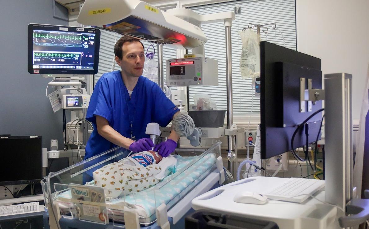
A quick, noninvasive eye scan performed on very premature infants in the NICU may predict how their brains will develop by age 2, according to a Duke University School of Medicine study published Wednesday, June 4 in JAMA Ophthalmology.
In a cohort of very preterm infants born at Duke, researchers found that the thickness of the retinal nerve fiber layer (RNFL), measured at approximately 36 weeks' postmenstrual age (roughly four weeks before a full-term due date), was significantly associated with cognitive, motor, and behavioral outcomes two years later. Infants with thicker RNFL had better developmental results.
"Because the retina is part of the central nervous system, it gives us a unique, noninvasive way to study the developing brain," said corresponding author Cynthia Toth, MD, a professor of ophthalmology at Duke. "Using a quick, safe eye scan, we may be able to identify infants at higher risk for developmental delays much earlier."
The scan uses optical coherence tomography (OCT), a handheld device that captures detailed retinal images with safe infrared light. It piggybacks on routine retinopathy of prematurity exams already performed in the NICU, adding no extra procedure for the infant.
The significance: very preterm infants face up to 50 percent higher risk of developmental challenges affecting movement, learning, language, and behavior, according to the researchers. Many of those challenges go unrecognized until later in infancy or early childhood, narrowing the window for early intervention.
Lead author Kathryn E. Gustafson, PhD, a professor in psychiatry and behavioral sciences at Duke, said the work moves clinicians closer to identifying which children need extra support much earlier in life. Co-author C. Michael Cotten, MD, MHS, a professor of pediatrics, said earlier insight into a baby's developmental trajectory could help care teams tailor follow-up and connect families with supportive therapies when they can have the greatest impact.
The findings emerge from BabySTEPS, an NIH-funded study that has been running at Duke since 2016. The long-running program uses bedside OCT imaging to identify early signs of retinal disease and indicators of neurological development in preterm infants. The research remains ongoing, though no specific timeline for clinical translation has been announced.